Description
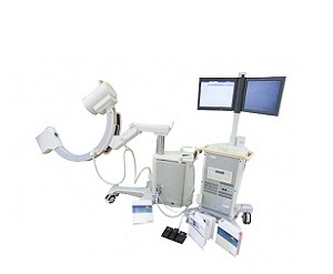
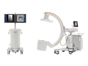
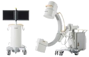
Philips BV Endura C-Arm X-Ray
Introducing the Philips BV Endura C-Arm X-Ray for sale. Confidently perform
open and minimally-invasive surgeries across multiple clinical specialties
including orthopedic, pain management and peripheral vascular procedures.
The BV Endura enables quick positioning, easy patient access,
and superb quality images to enhance workflow and decision making.
Philips BV Endura C-Arm X-Ray Features
Perfect for general fluoroscopy and vascular specialization,
the BV Endura helps you clearly view dynamic images in surgery.
Designed to improve your workflow, this versatile system has
many beneficial characteristics.
Positioned as a mid-level performer, the BV Endura builds on t
he strengths of the BV Libra by broadening vascular capabilities.
Our bigger 12” image intensifier provides a larger
coverage for improved anatomical orientation.
Extended C-Arm rotation – up to 135° – to provide you with
projections necessary for most vascular procedures.
Streamline vascular workflow
Philips BV Endura C-Arm X-Ray offers a wide range of vascular options with
different fluoroscopy and exposure frame rates and optional acquisition modes,
like C0², to enhance visualization during demanding vascular procedures.
The single user concept allows clinicians to use a footswitch and remote
control to control various imaging functions from the table.:
Subtraction, Trace, Roadmap, or Masking.
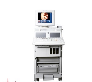
Optimally designed, the MobileView Station’s ultra-compact size makes it easy
to move around the tight confines of the OR. And its small footprint
ensures it can be brought as close as possible to the operating table.
At the MobileView Station the operator can manually enter patient
demographics or retrieve a work-list via the hospital network.
Monitors are turned toward the physician at any angle at the start
of the procedure and back to the operator at the end to process the images.
Processed images are immediately sent to PACS
Two 18” monitors ensure high quality image viewing
Monitor height can be quickly adjusted to physician preference
Philips BV Endura C-Arm X-Ray Specifications
Image intensifier type: Triple mode 9″ HRC / Triple mode 12″
>Nominal II formats
12″, 9″, and 7″
9″, 7″, and 5″
Entrance screen: Cesium Iodine
Grid type: Circular, carbon fiber; 60 lines/cm Ratio = 1:10 SID = 100 cm
TV camera type: CCD; high resolution 1K
Image rotation: Digital, live and on LIH
Image reversal: Yes
Mirror up/down: Digital, live and on LIH
Mirror left/right: Digital, live and on LIH
Automatic anatomical measuring field: Yes with ‘BodySmart’
Generator type: DC converter, micro-processor controlled
Max. generator output: 3.15 kW
Max. X-ray tube voltage: 110 kV
Max. X-ray tube current: 30 mA
Tube type: Fixed anode
Nominal focal spot values (IEC 336): 0.6 IEC and 1.4 IEC
Nominal X-ray tube voltage: 110 kV
Maximum anode heat content: 35.5 kJ = 50 kHU
Anode cooling capacity 21.6 kJ/min. = 30.6 kHU/min.
Maximum housing heat content: 840 kJ = 1200 kHU
Inherent filtration: 3.0 mm Al eq.
Additional filtration: 1.0 mm Al eq. + 0.1 mm Cu
kV range: 40 – 110 kV
mA range for Low Dose Fluoroscopy mode:0.10 – 3.00 mA (up to 7.20 mA during Auto High Penetration)
mA range for High Definition Fluoroscopy mode: 0.24 – 7. 20 mA
Shutters: Two independent lead shutters with steel wedge: shutters can
be coupled for rotation and translation, or moved individually for asymmetric collimation
Automatic Shutter Positioning: Automatic shutter placement based on image content
Shutter material: 3 mm Pb
Wedge material: 0.2 to 2.5 mm stainless steel
Adjustment: Stepless
Rotation: 360°
Minimal iris diameter: < 50 mm on II entrance
Position indication: On screen and also on LIH without radiation
Analog video out: 1 BNC connector left monitor
Digital Video out: 2 DVI connectors left and right
Video in: S-Video
USB storage: bmp format
Reviews
There are no reviews yet.